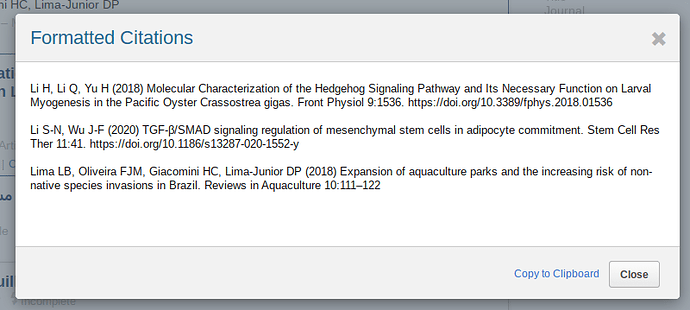
Thanks @vicente! I did run across a few others - it seems to come up most frequently in cases where one first author’s last name is on the shorter side (in bold).
Biferali B, Proietti D, Mozzetta C, Madaro L (2019) Fibro-Adipogenic Progenitors Cross-Talk in Skeletal Muscle: The Social Network. Front Physiol 10:1074.
Bi P, Ramirez-Martinez A, Li H, et al (2017) Control of muscle formation by the fusogenic micropeptide myomixer. Science 356:323–327.
Boucher J, Softic S, El Ouaamari A, et al (2016) Differential Roles of Insulin and IGF-1 Receptors in Adipose Tissue Development and Function. Diabetes 65:2201–2213.
Bou M, Montfort J, Le Cam A, et al (2017) Gene expression profile during proliferation and differentiation of rainbow trout adipocyte precursor cells. BMC Genomics 18:347.
Bour BA, O’Brien MA, Lockwood WL, et al (1995) Drosophila MEF2, a transcription factor that is essential for myogenesis. Genes Dev 9:730–741.
Duran BO da S, Dal-Pai-Silva M, Garcia de la Serrana D (2020) Rainbow trout slow myoblast cell culture as a model to study slow skeletal muscle, and the characterization of mir-133 and mir-499 families as a case study. J Exp Biol 223.:
Du SJ, Devoto SH, Westerfield M, Moon RT (1997) Positive and negative regulation of muscle cell identity by members of the hedgehog and TGF-beta gene families. J Cell Biol 139:145–156.
Du SJ, Dienhart M (2001) Gli2 mediation of Hedgehog signals in slow muscle induction in zebrafish. Differentiation 67:84–91.
Hepler C, Vishvanath L, Gupta RK (2017) Sorting out adipocyte precursors and their role in physiology and disease. Genes Dev 31:127–140.
He S, Salas-Vidal E, Rueb S, et al (2006) Genetic and transcriptome characterization of model zebrafish cell lines. Zebrafish 3:441–453.
Hesslein DGT, Fretz JA, Xi Y, et al (2009) Ebf1-dependent control of the osteoblast and adipocyte lineages. Bone 44:537–546.
Hopkins PM, Das S (2015) Regeneration in crustaceans. The natural history of the Crustacea 4:168–198
Ho SY, Goh CWP, Gan JY, et al (2014) Derivation and long-term culture of an embryonic stem cell-like line from zebrafish blastomeres under feeder-free condition. Zebrafish 11:407–420.
MacLea KS, Covi JA, Kim H-W, et al (2010) Myostatin from the American lobster, Homarus americanus: Cloning and effects of molting on expression in skeletal muscles. Comp Biochem Physiol A Mol Integr Physiol 157:328–337.
Ma J, Zeng L, Lu Y (2017) Penaeid shrimp cell culture and its applications. Rev Aquac 9:88–98.
Ma RC, Jacobs CT, Sharma P, et al (2018) Stereotypic generation of axial tenocytes from bipartite sclerotome domains in zebrafish. PLoS Genet 14:e1007775.
Maroto M, Reshef R, Münsterberg AE, et al (1997) Ectopic Pax-3 activates MyoD and Myf-5 expression in embryonic mesoderm and neural tissue. Cell 89:139–148.
Tanaka A, Woltjen K, Miyake K, et al (2013) Efficient and reproducible myogenic differentiation from human iPS cells: prospects for modeling Miyoshi Myopathy in vitro. PLoS One 8:e61540.
Tan X, Du SJ (2002) Differential expression of two MyoD genes in fast and slow muscles of gilthead seabream ( Sparus aurata). Dev Genes Evol 212:207–217.
Wei J, Glaves RS, Sellars MJ, et al (2016) Expression of the prospective mesoderm genes twist, snail, and mef2 in penaeid shrimp. Dev Genes Evol 226:317–324.
Weinberg ES, Allende ML, Kelly CS, et al (1996) Developmental regulation of zebrafish MyoD in wild-type, no tail and spadetail embryos. Development 122:271–280
Weintraub H (1993) The MyoD family and myogenesis: redundancy, networks, and thresholds. Cell 75:1241–1244.
Wei Q, Rong Y, Paterson BM (2007) Stereotypic founder cell patterning and embryonic muscle formation in Drosophila require nautilus (MyoD) gene function. Proc Natl Acad Sci U S A 104:5461–5466.
Yun Y-R, Won JE, Jeon E, et al (2010) Fibroblast growth factors: biology, function, and application for tissue regeneration. J Tissue Eng 2010:218142.
Yu T, Chua CK, Tay CY, et al (2013) A generic micropatterning platform to direct human mesenchymal stem cells from different origins towards myogenic differentiation. Macromol Biosci 13:799–807.

